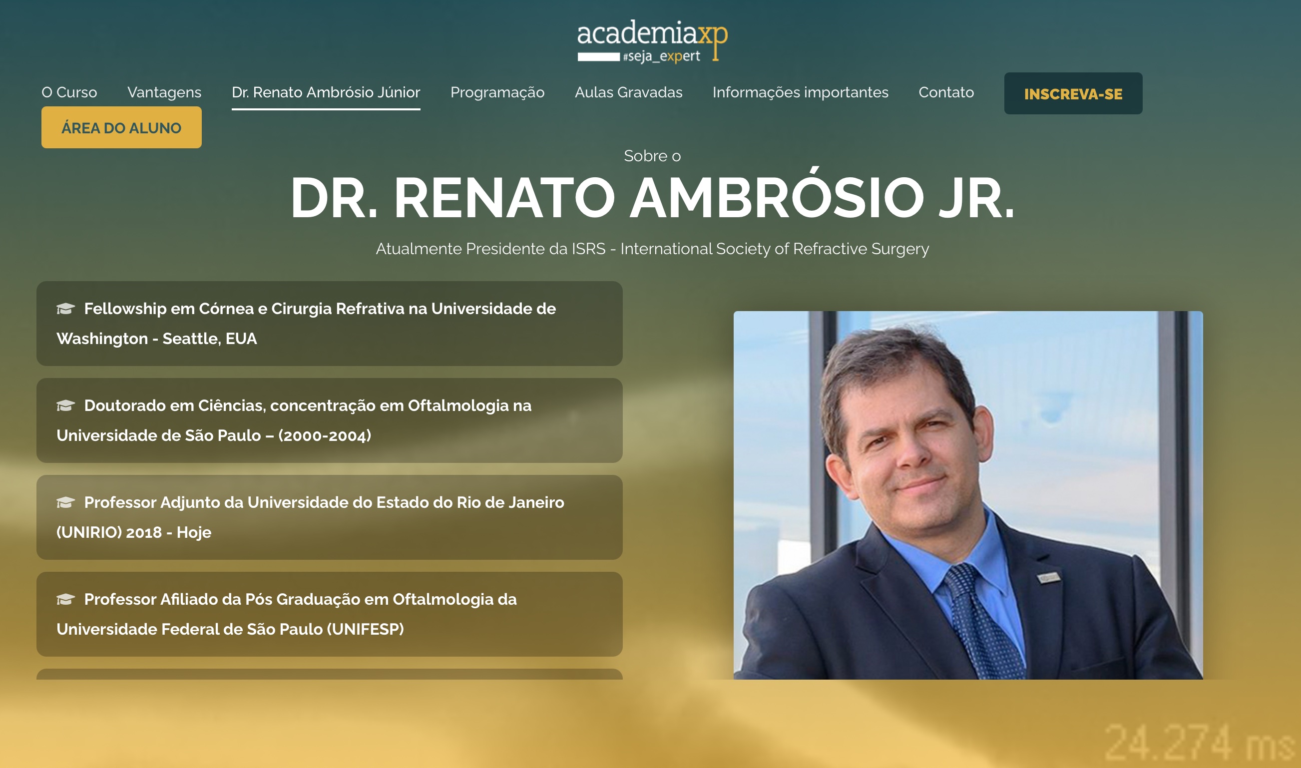
Dr. Ambrósio (Renato), you are our current president of the prestigious ISRS - The International Society of Refractive Surgery (www.isrs.org). You are among the most influential and famous specialists in refractive surgery, keratoconus, and cornea imaging worldwide. Please, tell us about yourself.
Thank you, Sebastian! I am happy to collaborate with Digital-Ophthalmology.net. Congratulations on this work.
I completed 49 years old in 2021. I was born in Rio de Janeiro, Brazil, where I graduated from Medical School in 1995. I belong to a family of many ophthalmologists. My father was among the pioneers of refractive surgery in Brasil in the early 1980s, my mother is a general ophthalmologist, my younger brother is a retina specialist, and my wife is also an ophthalmologist.
I did a residency and cornea fellowship in São Paulo and later a refractive surgery fellowship at the University of Washington in Seattle, USA. I was very fortunate to have great mentors, including Dr. Tadeu Cvintal and Dr. Steve Wilson. I early realized that our field is evolving fast and should focus on learning the foundations of ophthalmology, and of refractive surgery. So, I also learned from them how to think and to elaborate on the questions we should ask for progress. I consider myself a refractive surgeon-scientist.
You published many papers regarding the applications of Artificial Intelligence. What can Artificial Intelligence do, what you can´t do?
Last year we celebrated ten years of the BrAIN - Brazilian Artificial Intelligence Networking in Medicine. This is a multicentric group composed of researchers in Computer Science, Medicine and Physics from different regions of Brazil. I, João Marcelo Lyra and Aydano Machado started this strategic partnerships based on interdisciplinarity as a powerful tool for transformation.
Artificial intelligence (AI) is an important branch of computer science that has become increasingly part of our lives. We have it on smart assistants like Siri and Alexa, conversational bots, email spam filters, and even Netflix's recommendations. Soon, we will have self-driving cars and other robotic functions. While the world is changing fast with AI, this is here to help human life. Not to replace us, I believe. However, my concern is how we will adapt to the fact that some jobs and occupations will change. I understand that we as a society will evolve, and human care, creativity, and imagination will become more important for the next generation.
Do you think AI can help us detect corneal diseases, such as ectasia and keratoconus?
Absolutely, YES! First, we need to explain and clarify some terms. Keratoconus is the most important and more common form of corneal ectasia. From the Global 2015 consensus,1 the pathophysiology of corneal ectasia is related to biomechanical failure. A focal abnormality in corneal properties precipitates a cycle of decompensation, leading to localized thinning and steepening (bulging). The altered corneal shape generates myopia and astigmatism, which are measured as lower and higher order ocular wavefront aberrations. I may add that corneal properties are related to genetics.
While we are still not able to characterize genetic predisposition, we have multimodal refractive imaging.2,3 This includes Placido-disk based corneal topography, corneal and anterior segment tomography and segmental or layered tomography from Scheimpflug, OCT and digital very-high frequency ultrasound (dVHF-US), corneal biomechanics, ocular wavefront, ocular biometry, and cellular specular and confocal evaluations. This is a true revolution in evolution.
So, consider the amount of data that is translated into parameters, maps, and displays. Such an enormous amount of information can be processed very efficiently using AI. However, the major concept is to go beyond, not over, as we point out in the JRS Editorial.4 Beyond, not over considering the classic central corneal thickness and corneal front surface topography.5 And also, beyond the detection of mild disease, but into the characterization of the individual susceptibility to develop progressive ectasia.
You have also published papers that show that linking patient data and different imaging modalities like the Oculus Pentacam® and the CORVIS® allow amazing insights just in the early detection of corneal ectasia. In your opinion, what else is possible here, especially thinking about the inclusion of even more imaging data like anterior segment OCT or epithelial thickness maps based on it?
This is fundamental to understand the study questions so that you properly design the study and perform it. For example, as part of the Ph.D. thesis of Bernardo Lopes, we developed the PRFI (Pentacam Random Forest Index) that enhances the ability to detect ectasia susceptibility.6 The training dataset included the preoperative status of cases that developed post-LASIK ectasia. These cases are rare so that we need to be proactive in gathering these data.
For the TBI (Tomography-Biomechanical index), available in the ARV (Ambrósio, Roberts and Vinciguerra) display, we collected a large series of very asymmetric ectasia cases in joint work with Dr. Paolo Vinciguerra from Milan, Italy.7
AI needs proper training, which includes the measurements in well-defined populations. Training with internal validation should be carefully done. Also further external validation is a must.8 However, we are aware of some reports demonstrating lower accuracy of the TBI in certain populations,9 so that a much larger database was built from international centers to allow further training and optimization which allowed the second generation of the TBI.
You also are correct on the concept that more parameters can further improve. For example, in collaboration with Dr. Rohit Shetty’s group from Banglore, India, we demonstrated that OCT data, characterizing the epithelium and Bowman’s surface, could further augment accuracy.10
Furthermore, axial length and ocular wavefront may also improve the AI algorithms.
During the Corona pandemic, we all moved to increasingly use telemedicine. What are your experiences here? What is feasible and where is the personal contact to the patient missing, especially in corneal and refractive surgery?
Telemedicine is a reality. But we have limitations on the clinical exam, which we need to be wise on how to grow its use and how to optimize it. I have done some consultation calls which may work for some cases. But there is a need for exams and platforms such as the EyeLib are promising (https://mikajaki.com/). This should allow the patient be efficiently screened and send his data for the doctor evaluate in the consultation call.
In the last year, we have also been forced to do more of our training and teaching of students and residents online and digitally. I was also able to participate as a speaker in the EUCORNEA webinar series. What are your experiences with this? Is good teaching possible digitally?
Telemedicine and virtual conferences are growing in parallel. There is a strong need for education and training. I see the webinar as a great advance. We need, however, to find the proper balance. But I like the ability to participate in meetings without having to travel and be away from family. It also optimizes the time away from the clincial practice.
I have participated in many webinars and this is growing and getting better and better. Following the concept, I started my first online course with a MasterClass course on Multimodal Imaging. The first class was in Portuguese but soon, we will release an English version.
Soon you will also start your Masterclass in Multimodal Refractive Imaging online. Tell us a little more about it! How can we apply for it?
Thank you for asking. My goal is to create the course I always would like to take and offer it to help inspire and educate colleagues in the field I love. The address is already active and we have some content already available at https://renatoambrosiojr.academiaxp.com.br/eng/
Sounds fantastic! We will be happy to participate. So thank you very much for the interview, Renato!
Glad to collaborate with you. Thank you.
References:
1. Gomes JA, Tan D, Rapuano CJ, et al. Global consensus on keratoconus and ectatic diseases. Cornea 2015;34:359-69.
2. Ambrosio R, Jr. Multimodal imaging for refractive surgery: Quo vadis? Indian J Ophthalmol 2020;68:2647-9.
3. Salomao MQ, Hofling-Lima AL, Gomes Esporcatte LP, et al. Ectatic diseases. Exp Eye Res 2021;202:108347.
4. Ambrosio R, Jr., Randleman JB. Screening for ectasia risk: what are we screening for and how should we screen for it? J Refract Surg 2013;29:230-2.
5. Ambrosio R, Jr., Klyce SD, Wilson SE. Corneal topographic and pachymetric screening of keratorefractive patients. J Refract Surg 2003;19:24-9.
6. Lopes BT, Ramos IC, Salomao MQ, et al. Enhanced Tomographic Assessment to Detect Corneal Ectasia Based on Artificial Intelligence. Am J Ophthalmol 2018;195:223-32.
7. Ambrosio R, Jr., Lopes BT, Faria-Correia F, et al. Integration of Scheimpflug-Based Corneal Tomography and Biomechanical Assessments for Enhancing Ectasia Detection. J Refract Surg 2017;33:434-43.
8. Ferreira-Mendes J, Lopes BT, Faria-Correia F, Salomao MQ, Rodrigues-Barros S, Ambrosio R, Jr. Enhanced Ectasia Detection Using Corneal Tomography and Biomechanics. Am J Ophthalmol 2019;197:7-16.
9. Esporcatte LPG, Salomao MQ, Lopes BT, et al. Biomechanical diagnostics of the cornea. Eye Vis (Lond) 2020;7:9.
10. Chandapura R, Salomao MQ, Ambrosio R, Jr., Swarup R, Shetty R, Sinha Roy A. Bowman's topography for improved detection of early ectasia. J Biophotonics 2019;12:e201900126.


